Home » Without Label » Back Muscles Chart : Pin by Sarah Elledge on Lets Get PHIT and PHAT ladies ... : The superficial group, the deep group, and the intermediate group.
Back Muscles Chart : Pin by Sarah Elledge on Lets Get PHIT and PHAT ladies ... : The superficial group, the deep group, and the intermediate group.
Back Muscles Chart : Pin by Sarah Elledge on Lets Get PHIT and PHAT ladies ... : The superficial group, the deep group, and the intermediate group.. This allows you to pull your elbows back as far as possible, maximally stimulating the back muscles. There are three different muscle groups found in the back: The extrinsic (superficial) back muscles, which lie most superficially on the back. Flexes elbow and moves forearm. Our latest youtube film is ready to run.
Leaning back to straight vertical and all points in between. This helps concentrate more stress on the back muscles. It is the most superficial of all the back muscles. The soleus, the plantaris, and the gastrocnemius. Extends spine and trunk back.
Muscles Diagrams: Diagram of muscles and anatomy charts ... from i.pinimg.com Some of these muscles are quite large and cover broad areas. Muscle charts of the human body for your reference value these charts show the major superficial and deep muscles of the human body. Raises and rotates arm in all directions. Loss of control of the bowel or bladder and retention of urine may. Muscle injuries of the lower back are commonly caused by an improper lift, lifting while twisting, or a sudden movement or fall, which may cause lower back pain. There are three different muscle groups found in the back: The multifidus muscle keeps the back straight and stable. Your clients will thank you for it!
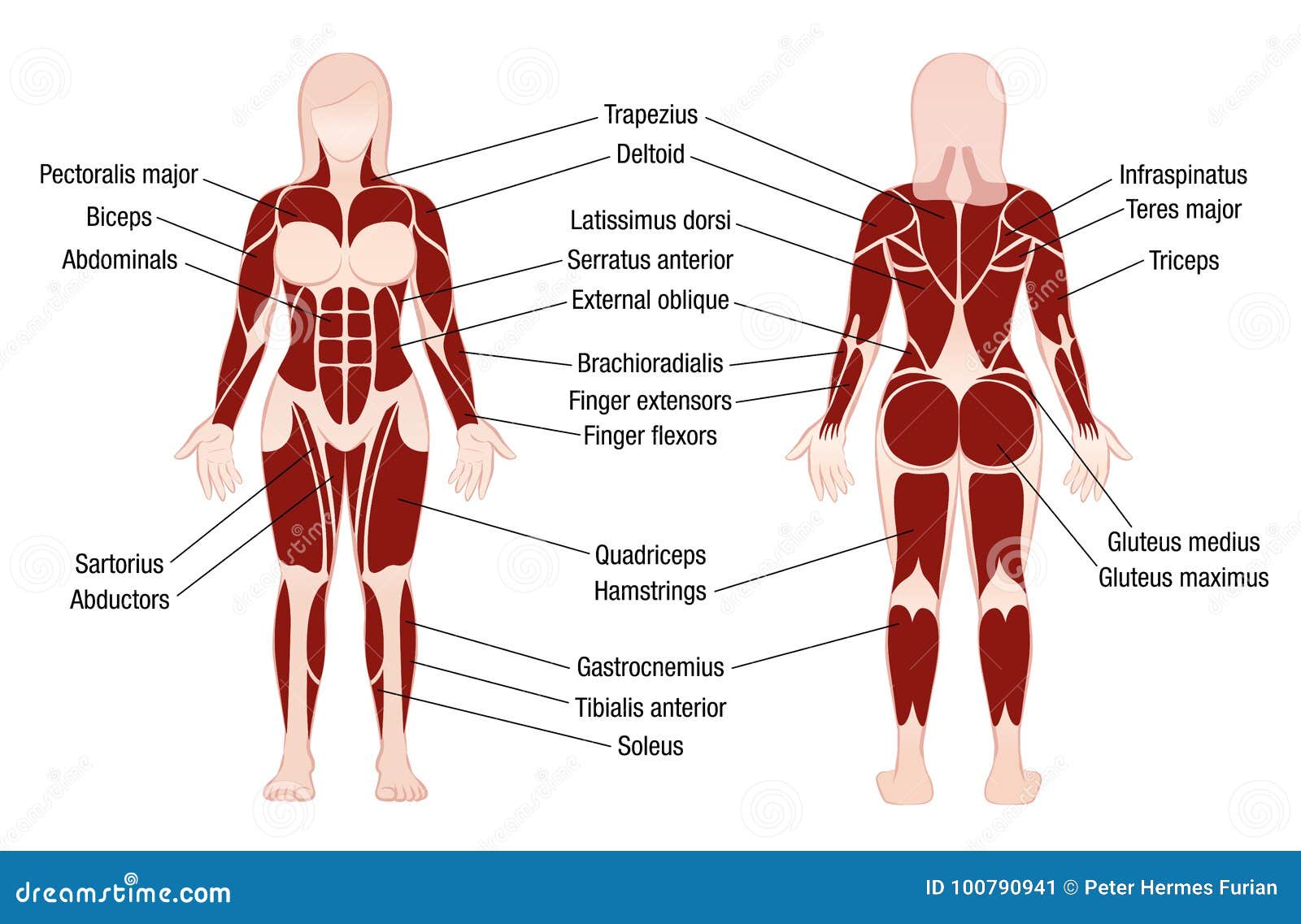
For images of the muscle, click on each link under location.
The deep back muscles, also called intrinsic or true back muscles, consist of four layers of muscles: Multifidus issues usually lead to other problems due to improper recruitment of other muscles to avoid pain. These muscles include the large paired muscles in the lower back, called erector spinae, which help hold up the spine, and gluteal muscles. Adductor magnus biceps femoris carpi flexor ulnaris deltoid. The trapezius and latissimus dorsi muscles connect the upper limb to the vertebral column. Back muscles, back muscle diagram. Keep your chest out and flexed throughout the move; Loss of control of the bowel or bladder and retention of urine may. We've created a free trigger point chart, which includes fybromyalgia treatment and reflexology information. Other muscles are small and cover much less space. Using both hands, pull up one knee and press it to your chest (b). 1) make midline incision along spines of vertebrae 2) extend from A strain can be an injury to a tendon attachment from muscle to bone.
The muscles of the back are a group of strong, paired muscles that lie on the posterior aspect of the trunk they provide movements of the spine, stability to the trunk, as well as the coordination between the movements of the limbs and the back muscles are divided into two large groups: Related posts of muscles of the lower back and hip diagram female back muscles diagram. Try these exercises to stretch and strengthen your back and supporting muscles. To download your free copy click the link. Musculoskeletal, shoulder & back back muscles, shoulder muscles.
Muscles Chart Description Muscular Body Woman Stock Vector ... from thumbs.dreamstime.com The muscles on each side form a trapezoid shape. Back muscles, back muscle diagram. The multifidus muscle keeps the back straight and stable. The deep back muscles, also called intrinsic or true back muscles, consist of four layers of muscles: The rhomboid muscle is activated as you bring and squeeze your scapula or shoulder blades back and together. Other muscles are small and cover much less space. Leaning back to straight vertical and all points in between. A strain can be an injury to a tendon attachment from muscle to bone.
The teres major is a small, yet important muscle within the back.
Some of the links in the post above are affiliate links.. Artery) p.134 accessory nerve p. Adductor magnus biceps femoris carpi flexor ulnaris deltoid. Both the deltoid and the trapezius are firmly attached to the spine of the scapula. Female back muscles diagram 12 photos of the female back muscles diagram diagram of female back muscles, female back muscles diagram, human muscles, diagram of female back muscles, female back muscles diagram. Brings hip away from body. This helps concentrate more stress on the back muscles. Muscle anatomy crossword answer key The back's muscles start at the top of the back (named the cervical vertebrae) and go to the tailbone (also named the coccyx). The fibres attach to the clavicle, acromion and the scapula spine. Creatine research more than a sports supplement read more…. The superior part of the appendicular skeleton that includes clavicle, scapula, and humerus, is attached to the axial skeleton that consists of skull. Muscle spasms (contraction or stiffening of the back muscles) muscles that feel tight;
An extremely strong tendon attached to the heel. The back's muscles start at the top of the back (named the cervical vertebrae) and go to the tailbone (also named the coccyx). We are pleased to provide you with the picture named anatomy of back muscles diagram.we hope this picture anatomy of back muscles diagram can help you study and research. We've created a free trigger point chart, which includes fybromyalgia treatment and reflexology information. Leaning back to straight vertical and all points in between.
Exercise Schedule for bodybuilders (Bodybuilder's Anatomy ... from cdn.shopify.com Muscles found in the superficial group include rhomboid major, rhomboid minor, levator scapulae, trapezius, latissimus dorsi. Superficial muscles of the back are located directly deep towards the skin along with superficial fascia.they are occasionally called the appendicular group as these muscles are mainly associated with activities of the appendicular skeleton. The superior part of the appendicular skeleton that includes clavicle, scapula, and humerus, is attached to the axial skeleton that consists of skull. 1) make midline incision along spines of vertebrae 2) extend from An extremely strong tendon attached to the heel. Extends spine and trunk back. We've created a free trigger point chart, which includes fybromyalgia treatment and reflexology information. Anatomy chart courtesy of fcit the latissimus dorsi muscles (also known as the lats) are the largest muscles of the back.
The trapezius and latissimus dorsi muscles connect the upper limb to the vertebral column.
Some of these muscles are quite large and cover broad areas. It is the most superficial of all the back muscles. Female back muscles diagram 12 photos of the female back muscles diagram diagram of female back muscles, female back muscles diagram, human muscles, diagram of female back muscles, female back muscles diagram. The vast majority of back problems improve on their own or with nonsurgical treatment. The rhomboid muscle is activated as you bring and squeeze your scapula or shoulder blades back and together. Superficial muscles of the back are located directly deep towards the skin along with superficial fascia.they are occasionally called the appendicular group as these muscles are mainly associated with activities of the appendicular skeleton. There are a few warning signs, however, that may indicate serious spinal problems. Muscle injuries of the lower back are commonly caused by an improper lift, lifting while twisting, or a sudden movement or fall, which may cause lower back pain. It is attached to the calcaneus and is pulled by 3 flexor muscles: Related posts of muscles of the lower back and hip diagram female back muscles diagram. Lie on your back with your knees bent and your feet flat on the floor (a). The superior part of the appendicular skeleton that includes clavicle, scapula, and humerus, is attached to the axial skeleton that consists of skull. Keep your torso upright and a slight arch in your back as you fully extend your arms at the top.